Immediate vs Delayed Implants: A Practical Guide to Placement
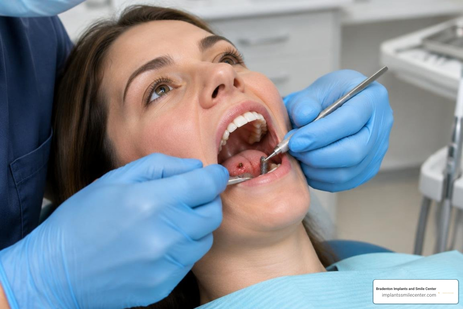
Understanding Immediate Placement Implants and Delayed Protocols
The timing of implant placement is classified based on the interval between tooth extraction and implant placement. The International Team for Implantology (ITI) categorizes these protocols into distinct types:
Type 1 (Immediate Placement): The implant is inserted directly into the fresh extraction socket during the same surgical appointment as tooth removal.
Type 2 (Early Placement with Soft Tissue Healing): The implant is placed 4 to 8 weeks after extraction to allow clear soft tissue resolution and closure over the socket.
Type 3 (Early Placement with Partial Bone Healing): The implant is placed 12 to 16 weeks post-extraction, providing time for substantial bone filling inside the socket.
Type 4 (Delayed/Late Placement): The implant is installed into a fully healed alveolar ridge, typically 6 months or more after extraction or ridge augmentation.
Choosing between immediate placement and delayed protocols involves balancing surgical efficiency against anatomical predictability. For a thorough walkthrough of surgical phases, review our comprehensive dental implant procedure guide.
Placement Protocol | Typical Healing Timeline | Required Surgical Visits | Primary Clinical Risk | Best Clinical Indication |
|---|---|---|---|---|
Immediate (Type 1) | 3–4 months to final restoration | 1–2 visits | Mid-facial soft tissue recession; loss of primary stability | Intact extraction socket, thick biotype, apical bone support |
Early (Type 2 / 3) | 5–7 months to final restoration | 2–3 visits | Soft tissue collapse over unhealed bone defect | Resolved soft tissue infection; minor wall defects |
Delayed (Type 4) | 6–10+ months to final restoration | 2–4 visits | Extended treatment time; alveolar ridge volume resorption | Extensive bone destruction, active purulence, thin biotype |
Surveys and clinical reviews indicate that patient satisfaction is high across all protocols when expectations are properly managed. Patients frequently prefer immediate placement due to fewer surgical appointments and reduced total treatment time. However, biological suitability must take priority over convenience.
Clinical Prerequisites for Immediate Placement Implants
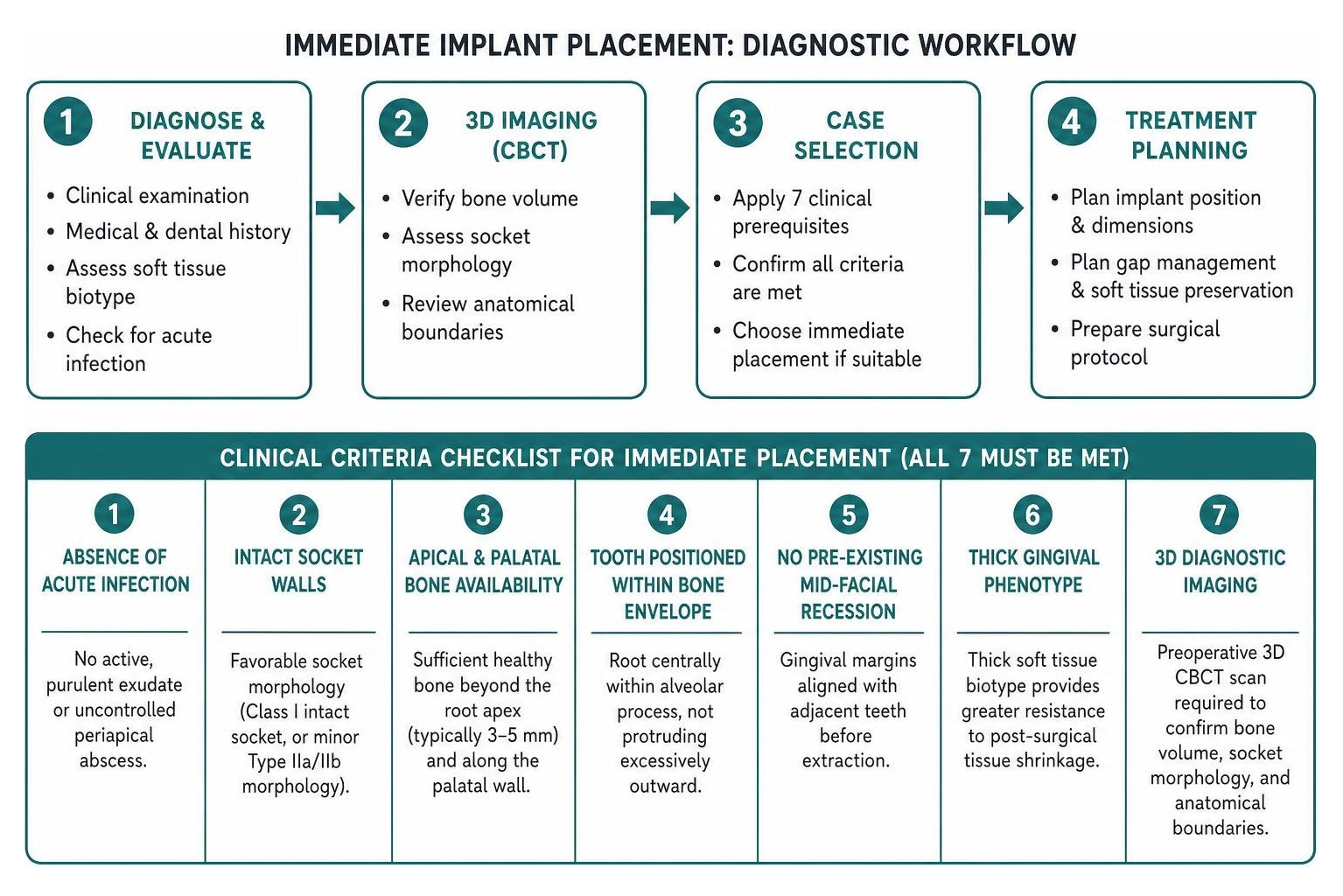
Immediate placement is not a default technique for every extraction. Predictable outcomes require a strict set of diagnostic criteria. The EAO Position Paper on tissue stability and case selection (Cosyn & Blanco, 2023) highlights seven core prerequisites that must be met simultaneously:
Absence of acute infection: No active, purulent exudate or uncontrolled periapical abscess at the surgical site.
Intact socket walls: Favorable extraction socket morphology (Class I intact socket, or minor Type IIa/IIb morphology).
Apical and palatal bone availability: Sufficient healthy native bone beyond the root apex (typically 3–5 mm) and along the palatal wall to anchor the implant.
Tooth positioned within the bone envelope: The native tooth root must sit centrally within the alveolar process, not protruding excessively outward.
No pre-existing mid-facial mucosal recession: The gingival margins must align with adjacent teeth before extraction.
Thick gingival phenotype: A thick soft tissue biotype provides greater resistance to post-surgical tissue shrinkage.
Three-dimensional diagnostic imaging: A preoperative 3D cone-beam computed tomography (CBCT) scan is required to verify bone volume, socket morphology, and anatomical boundaries.
Indications for Delayed and Staged Implant Placement
When local conditions do not meet these parameters, a staged or delayed protocol provides a safer pathway to long-term stability. Indications for delayed placement include:
Severe alveolar wall destruction: Complete loss or major dehiscence of the buccal bone plate.
Active, acute infection: Uncontrolled periapical or periodontal abscesses with extensive inflammation.
Insufficient native bone for primary stability: Inability to achieve primary mechanical anchorage without risking nerve impingement or maxillary sinus perforation.
Thin periodontal biotype with high esthetic risk: A thin, delicate gingival architecture prone to mid-facial recession in the smile zone.
In these situations, performing extraction socket preservation or guided bone regeneration first allows native bone and soft tissue to regenerate before implant placement. To explore how we customize these stages for complex anatomies, see our guide on developing a personalized dental implant plan.
Biological Healing and Osseointegration Dynamics
The biological processes governing healing around immediate implants differ substantially from those in healed ridges. When a natural tooth is extracted, the specialized bone lining the socket wall—known as the bundle bone—loses its functional stimulation via the periodontal ligament. Because bundle bone is tooth-dependent and predominantly vascularized by the periodontal ligament, it undergoes natural resorption during the initial weeks following extraction.
Immediate implant placement does not prevent bundle bone resorption. Even when an implant fills part of the socket, the outer buccal plate remodels. The biological response involves two overlapping phases of implant stability:
Primary (Mechanical) Stability: The mechanical friction achieved by engaging the implant threads into the native apical and palatal bone during surgery.
Secondary (Biological) Stability: The formation of new bone through osteogenesis directly on the implant surface, known as osseointegration.
As demonstrated in a comprehensive scoping review on biological mechanisms of implant placement timing, the transition from mechanical stability to biological osseointegration occurs between weeks 2 and 4. During this dip in total stability, cellular remodeling replaces mechanical friction with new woven bone.
Researchers analyze inflammatory markers and enzymes in peri-implant crevicular fluid to assess early wound healing kinetics. While bone remodeling pathways remain similar, immediate sites require careful gap management to prevent epithelial downgrowth along the implant collar.
Osseointegration Timelines and Marginal Bone Level Stability
Bone-to-implant contact (BIC) percentages in properly stabilized immediate implants match those achieved in delayed protocols over long-term observation. However, marginal bone level (MBL) changes during the first year require attention.
In delayed placement, the implant is surrounded entirely by mature, remodeled lamellar bone. In immediate placement, the coronal portion of the implant sits within a healing socket where bone remodeling occurs simultaneously with osseointegration. For an in-depth reference on how these biological stages ensure structural permanence, consult our complete guide to dental implants.
Long-term clinical trials show that when strict protocols are maintained, marginal bone level changes around immediate implants stabilize predictably after the initial remodeling phase, maintaining robust crestal bone support under functional loading.
The Jumping Distance and Socket Remodeling Dynamics
The space between the outer surface of the implant and the inner surface of the extraction socket walls is known as the horizontal gap or jumping distance.
Historically, clinicians believed that gaps smaller than 1.5 to 2 mm would heal spontaneously through blood clot organization alone. Modern clinical evidence shows that intentionally creating and grafting a horizontal buccal gap (>2 mm) produces superior ridge contour preservation.
By positioning the implant toward the palatal or lingual aspect of the socket, the clinician achieves two objectives:
It anchors the implant in dense native bone away from the thin buccal plate.
It leaves a wide buccal gap to receive bone graft material.
Preserving the periosteal vascular supply through a flapless extraction approach keeps the supraperiosteal blood vessels intact, minimizing post-extraction ridge resorption.
Surgical Execution, Primary Stability, and Gap Grafting
Achieving predictable outcomes with immediate placement implants requires specialized instrumentation and surgical techniques. Atraumatic tooth extraction is the first critical step; sectioning multi-rooted teeth and using periotomes or dynamic magnetic devices protects the delicate socket walls from fracture.
Once the socket is debrided, the osteotomy is prepared along the palatal or lingual wall. Using fully tapered implant designs with aggressive, deep apical threads aids in engaging the native bone beyond the root apex, securing the fixture in under-prepared bone sites.
Achieving Primary Stability and Insertion Torque Thresholds
Primary stability prevents micro-movement of the implant during the initial healing phase. Micro-movements exceeding 50 to 150 microns can disrupt capillary formation, leading to fibrous tissue encapsulation rather than osseointegration.
Key benchmarks for stability include:
Insertion Torque: A target insertion torque between 35 Ncm and 45 Ncm is standard for immediate placement. In prospective case series evaluating fully tapered implants, insertion torques consistently reached or exceeded 45 Ncm, providing rigid initial anchorage.
Implant Stability Quotient (ISQ): Measured via Resonance Frequency Analysis (RFA), an ISQ value of 65 or higher confirms sufficient rigidity for progressive healing.
Achieving these stability values safely relies on precise surgical technique, ensuring patient comfort throughout the procedure. We prioritize a gentle approach, utilizing advanced protocols designed for a pain free implant procedure.
The Role of Bone Grafting and Gap-Filling Materials
Filling the horizontal buccal gap is essential to counteract post-extraction socket remodeling. Evidence-based protocols, such as the 10 Keys protocol for immediate placement success, establish that grafting the buccal void maintains long-term soft tissue support.
Slow-Resorption Xenografts: Deproteinized bovine bone mineral or porcine-derived matrix is preferred over rapidly resorbing autografts or allografts within the gap. Because xenografts resorb slowly, the graft particles act as a long-lasting scaffold that supports the overlying soft tissue and maintains facial contour.
Subepithelial Connective Tissue Grafts: When the buccal soft tissue biotype is thin (<1 mm), placing a connective tissue graft inside a buccal tunnel envelope thickens the peri-implant mucosa and reduces the risk of recession.
To learn more about material options and regenerative techniques, explore our detailed resource on bone grafting for implants.
Esthetic Outcomes, Complications, and Provisionalization
In the esthetic zone (the upper front teeth), technical execution determines the visual outcome. While immediate and delayed implants demonstrate comparable implant survival rates, immediate placement presents a higher risk of mid-facial soft tissue recession if executed without appropriate case selection.
A common complication in immediate placement is positioning the implant shoulder too far buccally. When placed too close to the thin outer bone plate, inevitable bone remodeling exposes the metal collar or creates a grayish hue through the gingiva. Placing the implant 3 to 4 mm apical to the planned future mucosal margin and offset palatally avoids this issue.
Minimizing Esthetic Risk in Immediate Placement Implants
To protect papilla height and maintain a natural gumline, clinicians evaluate several anatomical factors before surgery:
Buccal Plate Integrity and Thickness: A facial bone wall thicker than 1.0 mm is less prone to vertical height loss than a paper-thin wall.
Interproximal Bone Peaks: The bone height on adjacent natural teeth dictates the height of the interdental papillae. If interproximal bone is intact, papilla preservation is highly predictable.
Restorative-Driven Angulation: Using computer-guided surgical stents ensures the implant axis aligns with the planned screw access channel, preventing buccal angular deviations.
Patients interested in seeing real clinical results across varied anatomical conditions can explore our dental implants before and after gallery.
Immediate Provisionalization and Pink Esthetic Score (PES)
Immediate provisionalization involves attaching a non-functional temporary crown to the implant on the day of surgery. This differs from immediate loading, as the provisional crown is adjusted out of all centric and eccentric occlusal contacts to prevent biting forces from stressing the implant.
A clinical evaluation of immediate placement with immediate provisionalization documented high esthetic and clinical success using this technique. In a prospective series of 24 patients receiving fully tapered implants with immediate restorations, investigators reported a 100% survival rate at 12 months. The mean Pink Esthetic Score (PES)—which grades papillae, soft tissue level, contour, alveolar deficiency, and color—improved from 11.08 at baseline to 11.48 at one year, well above the clinical acceptability threshold of 9. Furthermore, mean Marginal Bone Levels stabilized favorably from 2.44 mm to 2.16 mm.
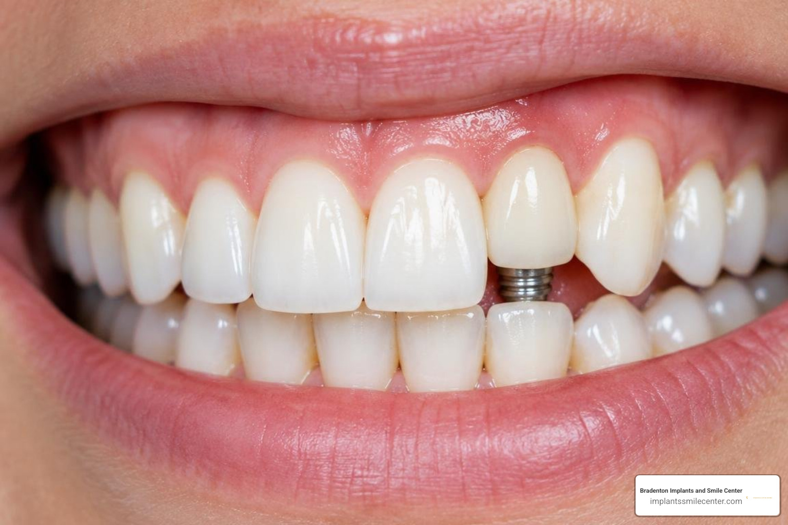
The immediate provisional crown acts as a prosthetic scaffold, supporting the severed gingival fibers and maintaining the natural emergence profile during healing. For candidates seeking to replace an entire arch efficiently, similar restorative principles apply to same day teeth options.
Frequently Asked Questions About Dental Implant Placement Timing
What are the main benefits of immediate placement implants?
The primary benefits include fewer surgical visits, a shorter overall treatment timeline, preservation of existing soft tissue contours, and the ability to leave the office with a fixed provisional crown when initial stability is favorable.
How do survival rates compare between immediate and delayed implants?
Both approaches demonstrate comparable long-term survival rates. Systematic reviews report a two-year survival rate exceeding 98% for immediate implants, with annual failure rates below 1%. While delayed placement shows slightly more consistent survival across compromised bone sites (98.3% to 100%), immediate placement achieves matching outcomes when strict selection criteria are followed.
When should an immediate implant procedure be aborted?
An immediate placement procedure should be converted to a staged approach if:
Insertion torque fails to reach the minimum threshold (e.g., <35 Ncm), indicating insufficient primary stability.
The buccal plate fractures or exhibits unexpected, extensive dehiscence during extraction.
Uncontrolled purulent infection is discovered within the apical socket space.
Anatomical constraints prevent leaving a minimum 2 mm horizontal gap between the implant and the buccal bone plate.
Conclusion
Choosing between immediate and delayed implant placement depends on accurate diagnostics, socket anatomy, and patient-specific esthetic risk factors. Immediate placement provides an efficient, tissue-preserving solution when anatomical prerequisites are met. When bone or soft tissue is compromised, staged and delayed protocols offer the stability needed for lasting results.
At Bradenton Implants & Smile Center, located at 7270 55th Ave E, Bradenton, FL 34203, we utilize high-resolution 3D CBCT imaging, guided surgical planning, and biological grafting protocols to tailor every procedure to your unique anatomy. If you are considering tooth replacement, schedule a consultation for advanced dental implants with our team to find the most predictable treatment path for your smile.
Scientific References
Cosyn J, Blanco J. "EAO Position Paper: Immediate Implant Placement: Managing Hard and Soft Tissue Stability from Diagnosis to Prosthetic Treatment.." The International journal of prosthodontics, 2023. PMID 37921675.