Dental Bone Graft Healing Stages and Pictures to Put Your Mind at Ease
What Bone Graft Healing Pictures Can Tell You
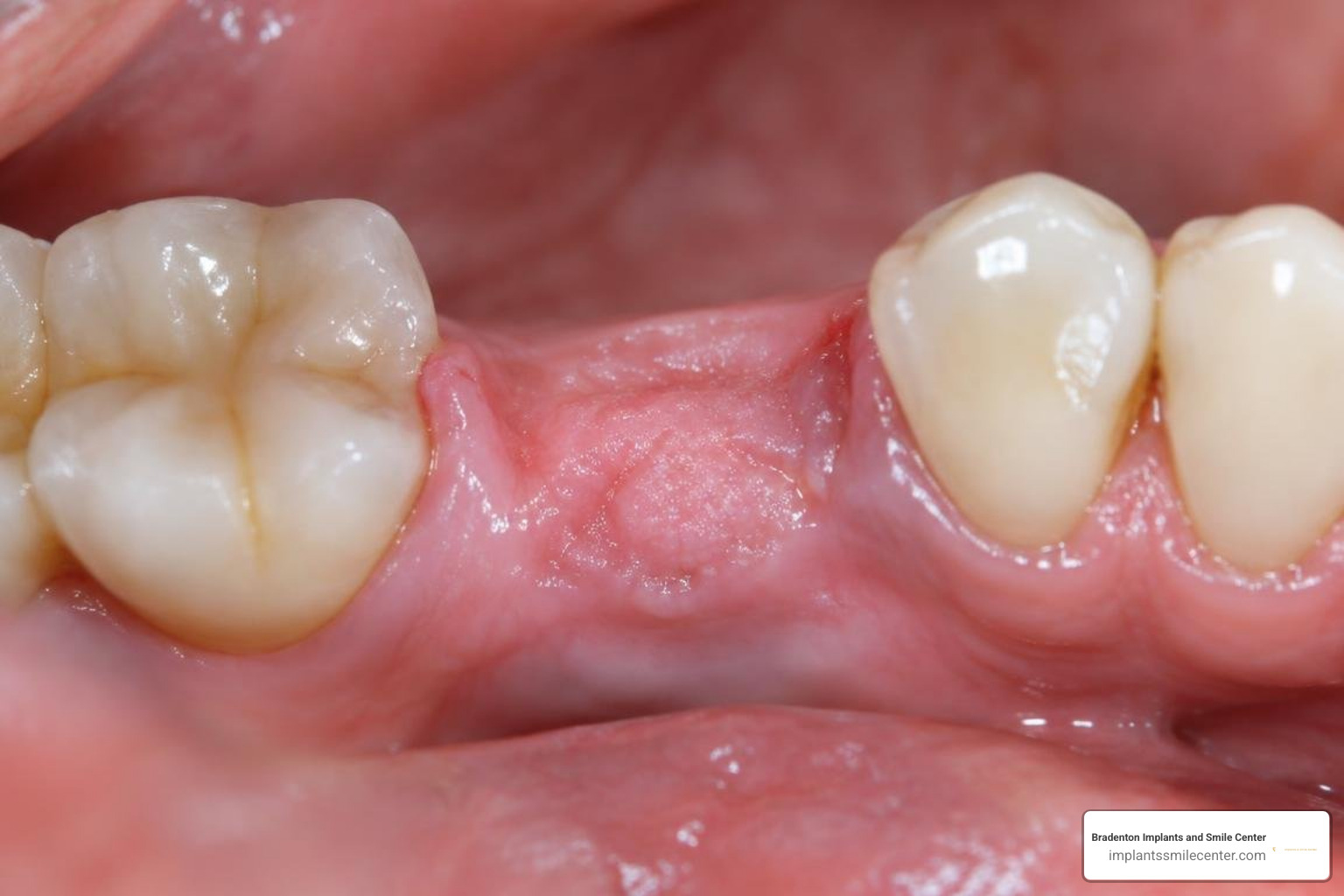
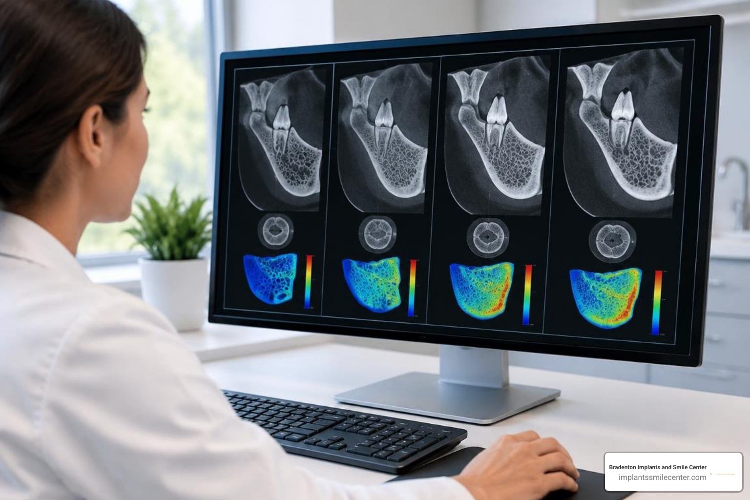
Bone graft healing stages pictures show a predictable pattern of tissue recovery: the surface gum tissue closes and matures first, while the underlying bone undergoes a complex multi-month cellular reconstruction. Immediately after surgery, dental radiographs show the physical placement of dense mineral particles. At two to four weeks, clinical intraoral photographs provide the clearest visual verification of soft-tissue closure, mucosal vascularization, and suture site health. Between three and six months, follow-up radiographs and three-dimensional CBCT scans illustrate internal mineralization and trabecular bridging. Reviewing pictures of bone graft healing stages at each interval helps you understand what is normal and what may require attention.
It is vital to recognize that a bright spot on an X-ray is not, by itself, proof that the graft has become new bone. Radiopacity merely confirms that mineralized particles are present within the socket or ridge defect. True biological integration requires living osteocytes to remodel that scaffolding into vital host bone. Your clinician carefully correlates photographic records, digital radiographic densities, and direct clinical palpation to evaluate structural healing.
I'm Dr. John Hegazin. With more than eight years of clinical practice in advanced restorative dentistry, bone augmentation, and implant surgery, I will guide you through what intraoral photographs and diagnostic radiographs show during each recovery phase.
Understanding the Biology of Dental Bone Regeneration
Dental bone regeneration is a highly organized, sequential cellular process. When placing particulate material into an extraction socket, lateral ridge defect, or maxillary sinus cavity, clinicians do not simply pack an inert filler into an empty space. Instead, surgical bone augmentation introduces a bioactive structural framework that initiates a coordinated physiological cascade driven by three foundational biological mechanisms:
Osteogenesis: The direct synthesis of new bone matrix by surviving, viable osteoblasts and pre-osteocytes contained within the donor material (a biological feature unique to autogenous bone harvested directly from the patient).
Osteoinduction: The biochemical recruitment, activation, and differentiation of pluripotential mesenchymal stem cells into functional bone-forming osteoblasts, stimulated by active bone morphogenetic proteins (BMPs) and growth factors present in specialized demineralized allografts.
Osteoconduction: The passive physical matrix or porous scaffolding that permits mechanical attachment, cell migration, capillary ingrowth, and new osteoid deposition across the surgical defect.
Reviewing the distinct types of dental bone grafts explains why particulate materials display variable resorption kinetics and differing radiographic densities over time.
Graft Material Type | Primary Biological Mechanism | Typical Resorption Rate | Clinical Strength |
|---|---|---|---|
Autograft (Patient's own bone) | Osteogenesis, Osteoinduction, Osteoconduction | Rapid to moderate | Gold standard for vital osteocytes |
Allograft (e.g., demineralized allograft fibers) | Osteoinduction, Osteoconduction | Moderate (3 to 6 months) | Excellent inductive signaling |
Xenograft (e.g., bovine-derived mineral matrix) | Osteoconduction | Very slow (6 to 24+ months) | Superior space maintenance and volume retention |
Alloplast (Synthetic bioceramics) | Osteoconduction | Variable depending on formulation | Predictable synthetic matrix |
The natural cellular processes that drive oral alveolar repair mirror general skeletal physiology. When skeletal or alveolar architecture suffers trauma, natural cellular bone repair mechanisms kick off immediately through localized inflammatory cascades, platelet aggregation, and specialized cell recruitment.
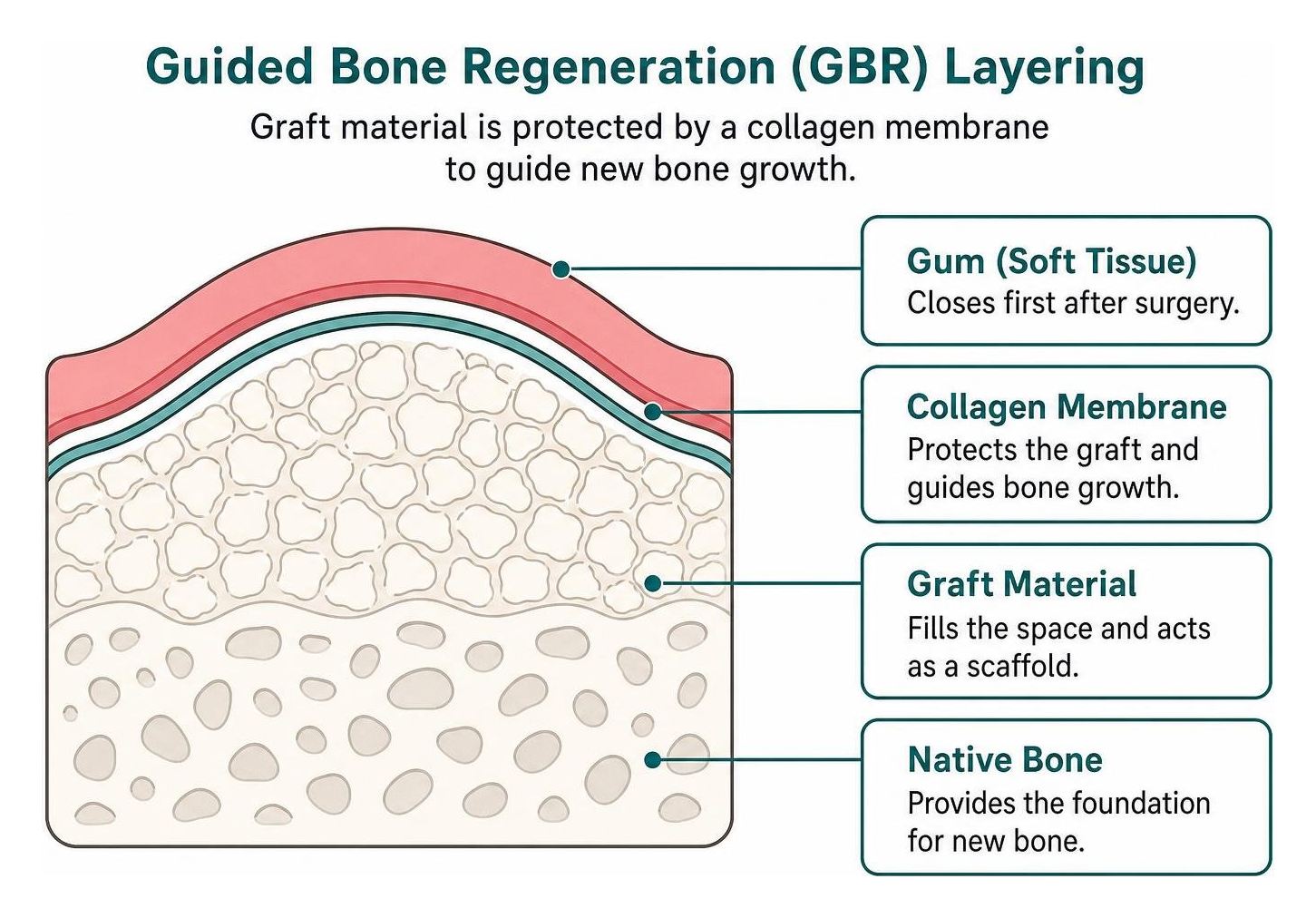
In guided bone regeneration (GBR), isolating the regenerative space is vital to overall success. Clinicians place a biocompatible, resorbable collagen barrier membrane over the grafted matrix. This physical barrier blocks rapidly proliferating epithelial cells and gingival fibroblasts from migrating into the bony void, reserving the space exclusively for slower-migrating osteogenic cells. Precise full-thickness flap management, tension-free periosteal releasing incisions, and stable primary closure using high-tensile monofilament sutures ensure the underlying graft remains stable, well-vascularized, and shielded from oral microflora throughout healing.
Visual Timeline: Bone Graft Healing Stages Pictures and Clinical Milestones
Documenting your recovery involves monitoring surface soft-tissue keratinization alongside deep radiographic remodeling. Achieving predictable bone grafting for implants requires sufficient mature, vascularized host bone before fixtures can support everyday masticatory forces.
Early Recovery: Bone Graft Healing Stages Pictures from Days 1 to 14
During the initial 24 to 48 hours following surgery, an organized fibrin blood clot stabilizes directly between the graft particles. Inflammatory cytokines recruit neutrophils and macrophages to clear surgical debris, followed rapidly by angioblasts that establish a fragile capillary network within the forming granulation tissue.
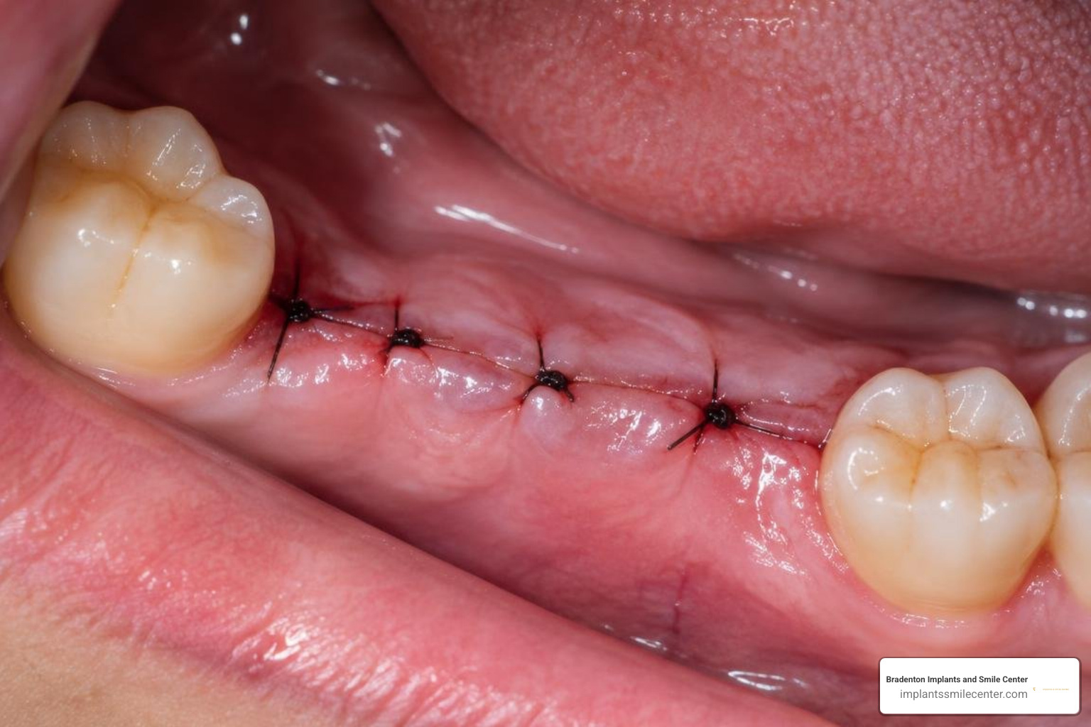
Between days 7 and 14, a provisional extracellular collagen matrix bridges the incision margins. Clinical intraoral photographs taken at this stage show the surgical gums shifting from acute post-operative erythema and mild edema to a stippled, pale-pink appearance. Tension-free sutures are evaluated and typically removed once the overlying epithelium establishes a tight seal over the underlying graft.
Bone Maturation: Pictures of Bone Graft Healing Stages at 3 to 6 Months
Between months 3 and 6, the provisional extracellular matrix undergoes progressive mineralization. Osteoblasts deposit disorganized woven bone along the graft scaffold, which osteoclasts subsequently remodel into dense lamellar trabeculae oriented along lines of functional stress.
In complex clinical scenarios—such as extensive 1-to-2 wall alveolar ridge defects measuring 10x8x5 mm following cyst enucleation—advanced multi-layer grafting techniques are especially valuable. Documented dual-layer graft clinical outcomes demonstrate that combining an internal osteoinductive demineralized allograft with a superficial slow-resorbing xenograft creates an optimal biological environment: the internal layer accelerates vital cellular osteogenesis while the outer mineral layer maintains ridge width and prevents contour collapse.
By 4 to 6 months, digital periapical radiographs and cross-sectional CBCT scans demonstrate continuous cortical bridging, absence of radiolucent gaps, and uniform bone fill, confirming the site is fully prepared for implant placement as detailed in our complete guide to dental implants.
Signs of Healthy Integration vs. Potential Complications
Distinguishing normal post-operative healing from early biological complications allows clinicians to intervene promptly and preserve the grafted site.
Normal recovery displays progressive reduction in localized tenderness within 3 to 5 days, steady pink mucosal adaptation, firmly retained sutures, and stable particulate containment beneath the flap. Conversely, persistent throbbing pain beyond one week, expanding dark red or purulent soft tissues, widespread particle leakage, or visible membrane exposure signal potential dehiscence or infection.
Systemic factors directly influence graft success. Uncontrolled diabetes, heavy nicotine use, immune suppression, and mechanical micro-motion disrupt capillary neovascularization, increasing the risk of graft failure. Diagnostic CBCT scans provide high-resolution cross-sectional validation, ensuring trabecular bridging without persistent radiolucency.
Post-Operative Care and Long-Term Maintenance Protocols
Protecting your regenerating bone graft requires diligent patient compliance at home paired with structured clinical monitoring:
Gentle Oral Hygiene: Avoid vigorous rinsing, spitting, or drinking through straws for the first two weeks. Do not touch or brush directly over the surgical site. Use prescribed antimicrobial chlorhexidine gluconate rinses twice daily as directed.
Dietary Modifications: Consume a soft, high-protein diet throughout the initial healing phase. Avoid crunchy, hard, or spicy foods, and chew exclusively on the opposite side of your mouth to prevent micro-motion.
Scheduled Clinical Reviews: Attend all planned follow-up appointments at 2 weeks, 4 weeks, 3 months, and 6 months post-surgery for clinical and radiographic evaluations.
Preventive Hygiene and Imaging: Maintain regular professional periodontal cleanings and annual low-dose digital radiographs to protect both the grafted ridge and the final restoration from peri-implant diseases, ensuring predictable long-term aesthetic stability as demonstrated in our dental implant before-and-after cases.
Frequently Asked Questions About Bone Graft Healing
How do I know if my dental bone graft is healing properly?
A properly healing bone graft exhibits a steady decline in post-operative swelling and discomfort within 3 to 7 days, healthy pale-pink gum tissue closure over the site, and complete absence of suppuration or foul odor. Radiographically, follow-up periapical images show stable mineral distribution without dark radiolucent halos or structural migration around the margins.
How long after a bone graft can I receive a dental implant?
Most routine socket preservation and localized ridge augmentation procedures require between 3 and 6 months of healing before the new bone achieves sufficient mineral density for implant placement. In select patients with excellent native bone volume and optimal primary stability, immediate implant placement can be performed simultaneously alongside particulate grafting.
What visual signs indicate a failing or infected bone graft?
Clinical signs of graft failure or localized infection include persistent deep-red or purple mucosal swelling, persistent or throbbing pain after the first week, active purulent drainage (pus), continuous shedding of loose bone granules into the mouth, premature soft-tissue dehiscence exposing the barrier membrane, and radiolucent dark bands visible around the graft on dental X-rays.
Conclusion
Rebuilding robust alveolar bone requires biological patience, but modern surgical techniques, bioactive biomaterials, and protective collagen membranes make the regeneration process exceptionally predictable. If you are planning tooth replacement or need to rebuild lost jawbone structure, schedule a consultation for comprehensive bone grafting and implant evaluations with our surgical team at Bradenton Implants & Smile Center.